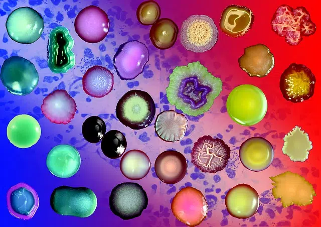
Staining is a technique used in laboratories.
The process and result of dyeing (giving a color to something) is called staining . The concept derives from the Latin word tinctĭo.
Importantly, the act and consequence of dyeing is also known as dyeing . In this way, while in colloquial language the idea of dyeing is usually used, in the field of chemistry and medicine the term dyeing is preferred.
Staining as a laboratory technique
In this sense, staining is a technique used in laboratories with the aim of optimizing the vision of what is observed through a microscope . Staining, in this way, consists of applying a dye to a substance or tissue to make it easier to detect and analyze.
With staining, it is possible to improve the definition of groups of cells or tissue fragments, to name a few options. Also, using special tinctures, the presence of certain substances or elements in a compound can be measured.

Thanks to the staining, the vision of what is observed through a microscope is optimized.
The in vivo process
When a living tissue is stained, one speaks of in vivo , supravital or vital staining. This allows us to observe chemical reactions or morphological characteristics of living tissues while the cells carry out their natural function. Generally, the objective that scientists seek through this type of staining is to bring to light certain data about the cytostructure (the conformation of the cell) that could not be observed in any other way, although it also serves to indicate the location of a specific chemical product, or of a reaction that occurs inside tissues or cells.
One of the main characteristics of in vivo staining is that dyes are usually used in highly dilute solutions, with concentration values ranging from 1:5,000 to 1:50,000. However, this does not always prevent the stain from being toxic to the body.
In vitro staining
We speak of in vitro staining to define the coloring of structures or cells that are no longer in their biological context . Various colorants are usually combined to obtain more detailed and precise results. When this is coupled with certain sample preparation and fixation protocols, science is able to produce consistent diagnoses.
The type of analysis desired for each particular case affects the steps necessary to carry out in vitro staining, and these are three: fixation , which consists of modifying the physicochemical properties of the proteins of a tissue or cell to preserve the maximum its form; permeabilization , to dissolve the cell membrane so that the dye can penetrate it; assembly , which seeks to increase the resistance of a sample so that it is not destroyed or loses its original structure throughout the process.
Gram's contributions
One of the best-known stains is the Gram stain , developed by Christian Gram , which allows bacteria to be visualized in clinical samples. Bacteria that react by turning purple are called Gram-positive bacteria , while those that turn pink are defined as Gram-negative bacteria .
Wright's stain , hematoxylin-eosin stain , and silver stain are other types of stains that can be used.
The counterstaining
There is also the concept of counterstaining , which refers to the application of a second stain to a certain preparation to make visible those parts that could not be stained with the first. Staining, on the other hand, can be indirect or direct according to the interaction of the dye with the fabric .
We can see an example of this technique when crystal violet (a group of compounds that are often used as dyes and pH indicators and is also called gentian violet or methyl violet ) is applied to a sample of bacteria for a Gram stain. since only the Gram positive ones are stained; For this reason, the application of safranin becomes necessary, which affects all cells and, therefore, allows Gram-negative cells to be identified.
