Using x-rays, an x-ray allows you to obtain an image of the inside of the body.
Radiography is the technique that, through the use of X-rays , allows obtaining an image of the inside of the body . The term is also used to name the photo generated with this technique.
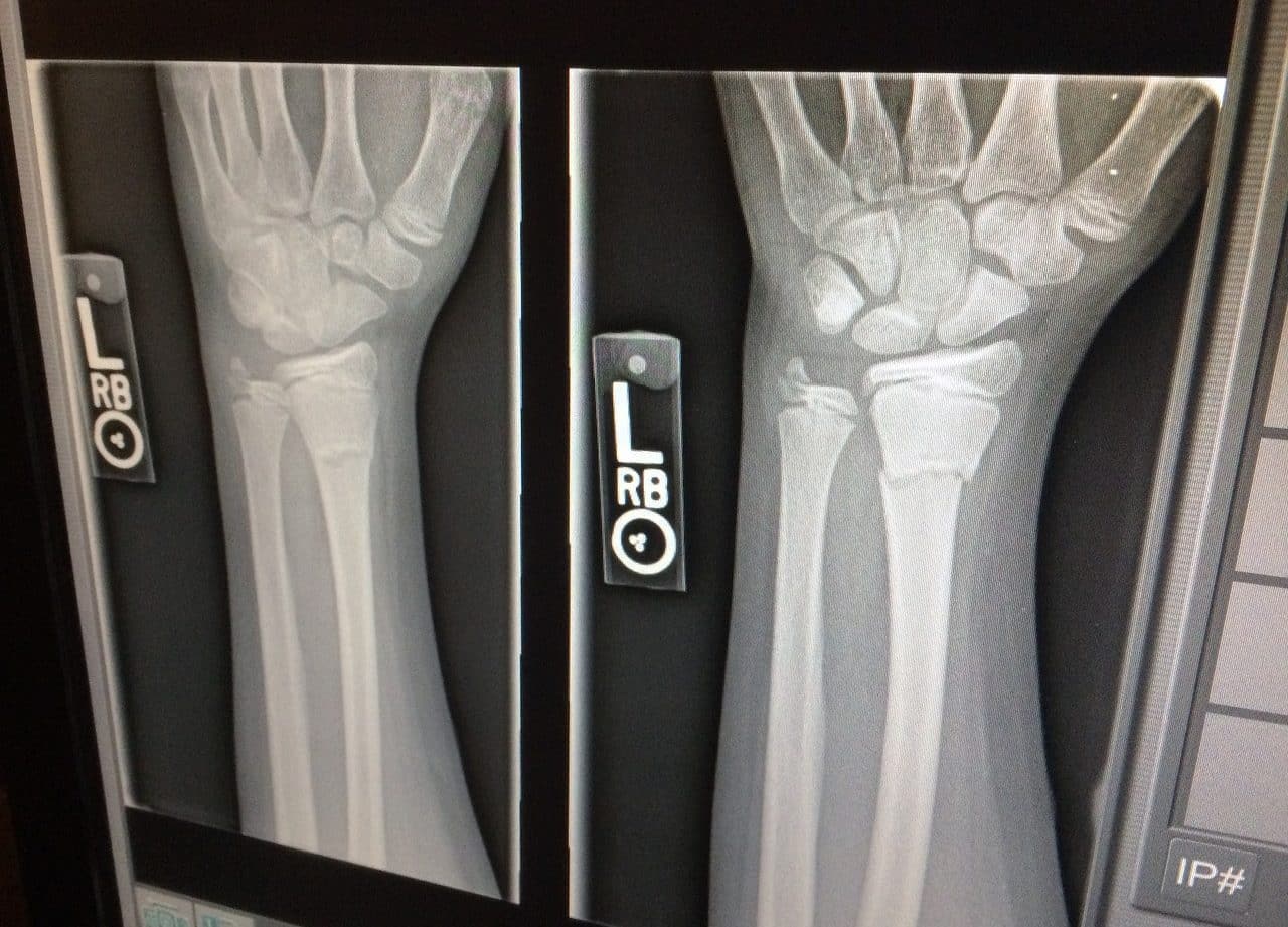
The procedure consists of exposing what you intend to photograph to a radiation source: that is, X-rays are emitted on the part of the body whose interior you wish to observe. X-rays have the ability to pass through soft tissues (organs, muscles, etc.), but not bones, which absorb the radiation. In this way, by placing a special detector behind the body, the X-rays generate the image . The bones are "engraved" in white and the rest of the internal components of the body, in different shades of gray according to density . The void, finally, remains black.
Thus, the x-ray is a photo that allows us to observe the bone components in white, on a black background. This helps a doctor to make different diagnoses according to the state of the bones .
Effects of x-ray on the body
It is important to note that, because the radiation doses to which the body is subjected are very low, x-ray is a safe procedure with minimal possibilities of causing damage to the body. Most experts assure that radiography offers benefits far greater than the risks it may entail.
Likewise, given that x-rays do not generate any perceptible effect on our body , if it were not for the machinery, the noise and the positions that the operators require us to maintain while taking the shot, it could be said that it is an equivalent procedure. than taking a conventional photograph . Despite being painless, the x-ray can leave us with some discomfort for a while due to posture and the inability to breathe normally.
These requirements, remaining still in a certain position and holding your breath for a few seconds, are typical of many of the most common types of x-rays, and their objective is to avoid blurred images, a common consequence if the body is in motion .
Before undergoing an x-ray, patients must meet certain requirements . For example, women should tell their doctor if they are pregnant or have an IUD. On the other hand, since metal objects can generate poor definition images, it is necessary to remove all accessories made of this material, such as jewelry, watches and belts; For added safety, it is recommended to use a hospital gown.
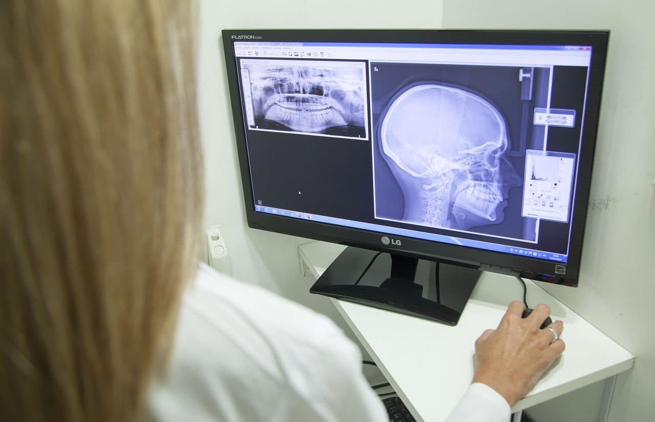
Doctors can make diagnoses thanks to x-rays.
Its usefulness
Among the most common types of x-rays are the following: abdominal ; of bone; chest; of the teeth; of a limb; of the hand; of the joints; of the neck; of the paranasal sinuses; of the skull; of the thoracic spine ; and the skeleton in general.
Radiography presents certain limitations, both physically and economically: safety protocols related to radiation doses cannot be ignored; As a testing method, it is very expensive; Any discontinuity that is not parallel to the radiation beam may be difficult to identify; The post-shot stage is extensive and involves various complementary procedures, such as image processing, drying and interpretation; and does not always offer reliable results.
Radiography beyond medicine
Beyond the medical level, the exhaustive analysis that is carried out on a topic is known as an x-ray.
For example: «The new chronicle published by the Argentine writer offers an excellent x-ray of life in the countryside» , «We have presented a very complete x-ray of the local economy so that the mayor can analyze which are the most convenient measures» .