The pelvis is made up of the coxal bones, the coccyx and the sacrum.
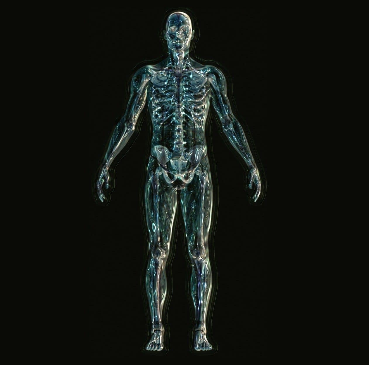
The pelvis is the body area of mammalian animals (including humans ) that is formed with the coxal bones , the coccyx and the sacrum . Located in the lower region in the case of the human body, it houses various organs of the reproductive system, the urinary bladder and the last section of the digestive tract.
With a funnel shape, it is possible to divide the pelvis into major and minor. The greater pelvis houses the organs of the abdomen , while the lesser pelvis contains the urinary bladder, genital viscera, rectum and anus.
The coxal bones, the sacrum and the coccyx make up the so-called bony pelvis . The region that is responsible for the articulation of the lower limbs, on the other hand, is known as the pelvic girdle .
About the pelvis
It is important to highlight that the man 's pelvis has several differences compared to the woman 's pelvis. In the female gender, the pelvis has a greater opening, is shorter and has thinner walls.
When a woman suffers from pelvic pain , on the other hand, it may be a symptom of endometriosis, ovarian cyst, an infection or an ectopic pregnancy, among other health disorders.
The posture and movement of the pelvis are very important in various dances and dances . The famous belly dance , also known as Arabic dance , is based largely on the movement that the dancer performs with her pelvis and the agitation she applies to said area.

Belly dancing consists of movements of the pelvis.
The fracture
A pelvic fracture is considered extremely serious since it can compromise the mobility of the hip, although it does not usually affect the stability of the pelvic ring. It usually occurs in patients who have suffered violent trauma to the lateral aspect and the greater trochanter. This type of fracture can cause damage to the pubic or anterior pillar, to the ischial or posterior pillar, to both or to the bottom of the cup (cavity of one bone into which the head of another enters).
All cases of pelvic fracture involve the acetabulum cavity; This can occur to a lesser degree (when the cup suffers a fissure) or completely (if the femoral head partially or completely sinks and is introduced into the pelvic cavity). Depending on the extent of the fracture, the damage to the joint varies, which may include the displacement of bone fragments.
To determine the magnitude of the pelvic fracture and the subsequent joint damage , a conventional x-ray is not enough, since in the resulting image it may be difficult to see the displacement of the ischial and pubic fragments. For this reason, two special projections become necessary:
- Alar : an x-ray that is made by rotating the pelvis at 45° towards the injured part, so that the iliac wing is located parallel to the plate and reveals the pubic pillar with the full extent of the fracture feature.
- Obturator or ischial : an x-ray is carried out with the pelvis rotated 45° in the opposite direction to the previous case, so that the ischial pillar, the obturator foramen and the fracture are completely visible.
Pelvic fracture is usually complicated by massive hemorrhages due to vascular damage to the obturator and iliac vessels. Furthermore, containing and reducing displaced fragments is very difficult. As a result of the involvement of the cup, it is very common for the hip to suffer from degenerative osteoarthritis ; This surely occurs when the bone damage to the joints is considerable and very frequently the mobility of said area is completely lost. The results of surgical interventions are uncertain since they are high- risk practices.