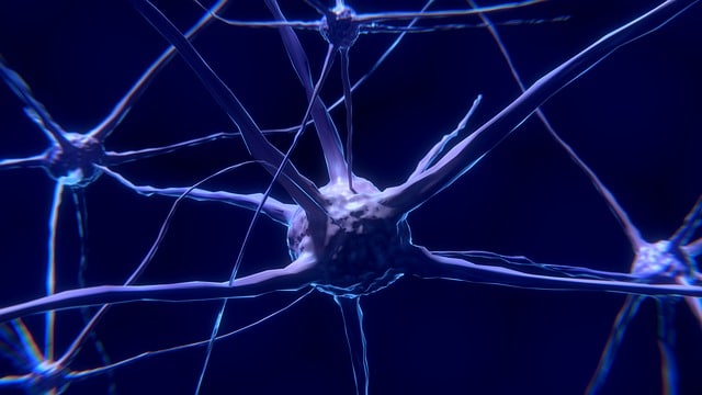
Microtubules are protein filaments found in cells.
The term microtubule comes from the English word microtubule . The concept is formed with the compositional element micro- (which refers to something "very small" ) and the noun tubule (a small tube that, in the field of anatomy , refers to an excretory duct).
protein filament
A protein filament found inside cells is called a microtubule. Microtubules are found in the cytoskeleton , in cilia and in other cellular organelles.
These microtubules originate from a structure called the microtubule organizing center . Depending on whether it is a prokaryotic cell or a eukaryotic cell, they have different characteristics.
The transfer of substances and the movement of organelles are some of the functions that microtubules have. They also intervene in the cell division processes ( meiosis and mitosis ).
Microtubules are also very important to give the cell its shape and to preserve its physiognomy. On the other hand, they function as transportation pathways for various molecules and vesicles.
Physical characteristics
Each microtubule is made up of protein polymers . Its outer diameter is around 25 nanometers, while its inner diameter barely exceeds 10 nanometers. They are considered heteropolymers of α- and β-tubulin , the latter being a family of globular-type proteins that also includes γ and that do not resemble almost any other protein. Its structural unit is dimers (macromolecular complexes that bring together two micromolecules, generally through non-covalent bonds), and these carry out polymerization into thirteen protofilaments, which can be seen on the sides as hollow cylinders.
For polymerization to take place, it is necessary that these dimers exist, but their concentration must be critical , which is minimal. By adding cores it is possible to speed up the process. Microtubules stand out mainly for their polarity: the polymerization of tubulin occurs by adding dimers on one side or both, joining the head of one with the tail of the other, and in this way rows of monomers are generated that give rise to a global polarity. Because all microtubule protofilaments are oriented in the same way, it is possible to identify a negative and a positive end, both formed by rings: the first, of α-tubulin and the second, of β-tubulin.
Bacterial microtubules
Although bacterial microtubules have a basic structure equivalent to that of eukaryotes, their diameter is smaller. Furthermore, in this case we should talk about bacterial tubulin proteins instead of tubulins , and use the letters A and B instead of α and β .
The microtubules of bacteria have protofilaments that are ordered and interact with each other almost as those of eukaryotes do. The last difference that we can point out is the number of protofilaments, since in eukaryotes there are thirteen, while in bacteria, only five.
Microtubules and cancer
It is interesting to note that knowledge about microtubules becomes especially relevant in the fight against cancer . There are drugs used in treatments that cause microtubules to not function correctly, which makes it difficult for cancer cells to multiply .
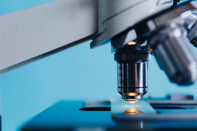
Some drugs manipulate microtubules to fight cancer.
The use of paclitaxel and other stabilizing drugs, in this framework, allows acting on specific regions of the microtubules. This makes it possible to hinder the progression of cancer.
Speaking specifically of paclitaxel, it is a drug discovered in 1968 at the Research Triangle Institute, when researchers Mansukh C. Wani and Monroe E. Wall managed to isolate a compound from the bark of the conifer tree called Taxus brevifolia and noticed that it acted against various types of tumors.