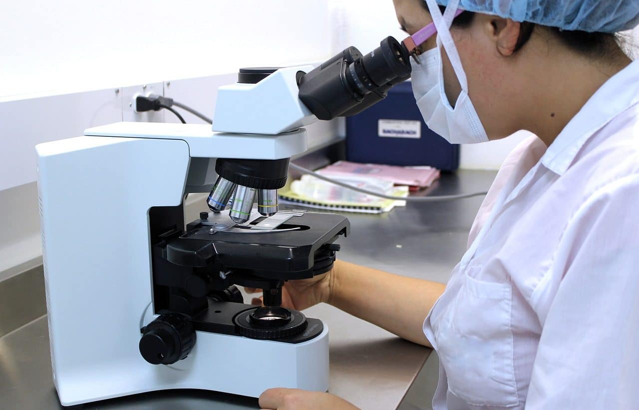
The microscope is very important in histology.
A microscope is a device that makes it possible to observe elements that are too small to be seen with the naked eye . The term comes from the Latin word microscopium.
The origins of the microscope date back to the end of the 16th century and are usually associated with tests carried out by the Dutchman Zacharias Janssen , a lens manufacturer. From the development of these instruments, science achieved great advances, such as the discovery of the existence of cells and the possibility of visualizing bacteria and other microorganisms.
History of the microscope
The approximate year to which Janssen 's invention of the microscope is related is 1590 . Almost a century later, in 1665 , the English physician William Harvey used this device to observe blood capillaries in the context of his research into blood circulation.
The British scientist Robert Hooke , for his part, was another of the pioneers in the use of the microscope; In his case, he documented his observation of a sheet of cork, in which he noticed the porosity of the material and the cavities in the shape of small cells that he called cells , the first dead cells to be observed. Shortly thereafter, Italian biologist and anatomist Marcello Malpighi used the microscope to look at living tissues and became the first scientist to detect living cells.
One of the most significant names in this framework is that of Anton van Leeuwenhoek , a Dutch merchant from the mid -17th century who made microscopes on his own and used them for the description of red blood cells, sperm, bacteria and protozoa. Despite not having scientific studies, this man became one of the founders of bacteriology . Although he decided not to reveal his techniques and methods, after his death the Royal Society of London for the Advancement of Natural Science acquired many of his devices.
The 18th century arrived and with it a series of advances that allowed the design of the microscope to be improved and varied. For example, Chris Neros and Flint Crown managed to create achromatic lenses , which were later improved by John Dollond . The outstanding studies of Isaac Newton and Leonhard Euler date from the same time.
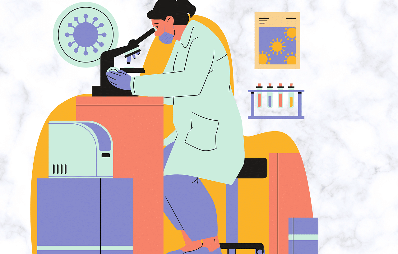
The analysis of viruses is possible thanks to microscopes.
Its development in the 19th century
During the 19th century it was discovered that the phenomena of refraction and dispersion could be modified by appropriately combining various optical media. This led to the launch of the best achromatic lenses ever created.
For the stability of the microscope, one of its fundamental properties, certain advances at a mechanical level were necessary. Another of the most important aspects is the number of increases , a feature that began at more than two hundred and currently exceeds one hundred thousand.
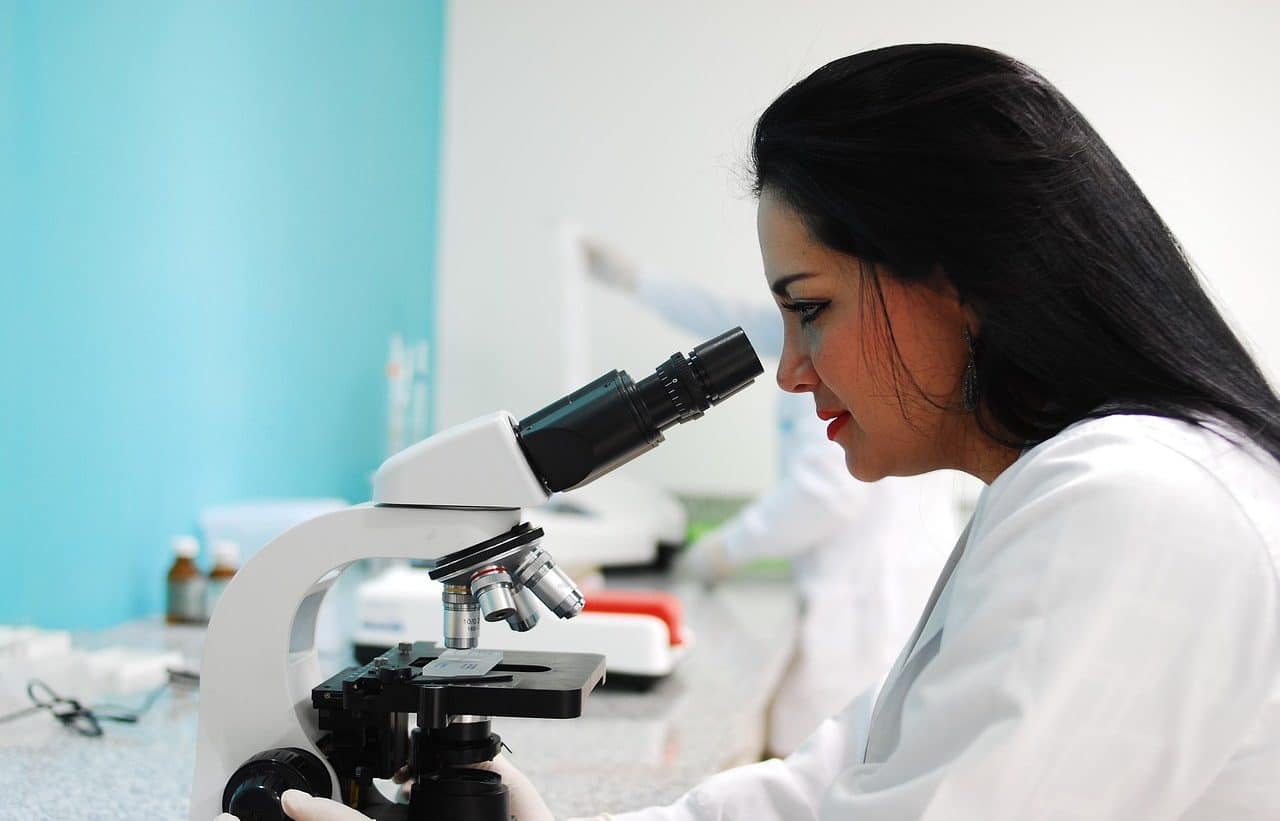
The study of the cell cycle is carried out with microscopes.
Types of microscope
There are various types of microscopes. The optical microscope is one that uses a system of lenses to magnify images, using visible light . In this group we find the simple microscope , which is the most basic instrument of this type: it has a single lens to increase the size of what is observed, as in the case of the magnifying glass.
The compound optical microscope , meanwhile, generates a magnified image thanks to the use of two or more optical systems. Each of these systems , which act successively, has one or more lenses.
The electron microscope , on the other hand, does not use visible light, but rather uses electron radiation . In this way the magnifications achieved are much higher than those offered by the optical microscope.
The most important difference between electron microscopy and optical microscopy, in short, is that the former uses electrons and the latter uses photons . The divergence in the wavelength of electrons and photons marks the possibility of achieving larger amplifications.
Its use
The microscope is key in biology, especially in the field of microbiology . This specialty is dedicated to the analysis of microorganisms.
In this context, the Petri dish acquires great relevance. It is a transparent container that is used in laboratories for cell culture, observation of microbes and seed germination, for example. The sample is placed inside (using agar or other culture medium) and then studied with a microscope.
In a general sense, it can be said that the microscope is used to enlarge the image of the sample, which in turn favors the distinction and separation of its components or constituent elements by making the details visible. Currently, microscopes usually also have lighting , another very important characteristic when it comes to promoting research.
There are countless discoveries made possible through the use of the microscope. One of the most important took place at the end of the 19th century , when the Spanish Santiago Ramón y Cajal began the development of the neuron doctrine .
This scientist, who received the Nobel Prize in Physiology and Medicine in 1906 , managed to observe, using staining techniques, the existence of functional and independent cells in the brain.