
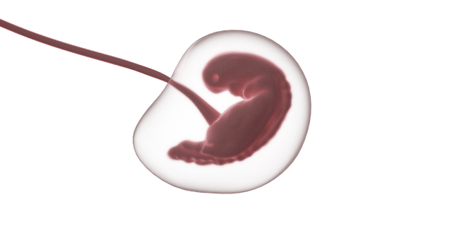
The embryoblast arises on the fourth day of embryonic development.
The embryoblast is a structure that is part of the blastocyst . It arises around the fourth day of embryonic development, coming from a mass of cells in the morula .
Originated in the morula
Once the zygote is formed after fertilization, successive divisions begin to develop that increase the number of cells and give rise to blastomeres (embryo cells). The blastomeres modify their shape and organize themselves until they become compact to promote intercellular communication.
When the divisions reach 32 blastomeres, the mass known as morula appears. The embryoblast arises from the inner cells of the morula and the trophoblast from the outer cells.
It is interesting to note that, of the 107 cells of the blastocyst, the majority correspond to the trophoblast: we are talking about 99 of them. Only eight cells , then, are from the embryoblast.
Layering
The embryoblast first divides into two layers and then transforms into three layers: the ectoderm , mesoderm , and endoderm . It is important to mention that each layer enables the development of different tissues and organs.
The respiratory system and the digestive system arise from the endoderm, for example. The mesoderm is the starting point of the heart and kidneys , among other organs, while the ectoderm allows the appearance of the peripheral nervous system , the central nervous system and other structures.
Division into two layers
The embryo takes several days to develop; When the eighth day arrives, and at the same time that the morula is fixed in the uterus, the first division of the embryoblast occurs: the layers called epiblast and hypoblast appear. The first is the superior. All its cells, which have a radial arrangement, are long and cylindrical in shape, are oriented in a single direction, towards the so-called embryonic pole .
In the internal part of these cells, the amniotic cavity is generated, which houses the fluid of the same name. With respect to the hypoblast, the lower layer, we can say that its cells have two orientations, including the abembryonic pole , they have a cubic shape and can be grouped into two layers. This stage comes to an end when the amnioblasts synthesize the amniotic fluid.
Division into three layers
This second division takes place after three weeks. At this point, the structure of the embryoblast is no longer spherical, but elongated, as if formed by two ovals . Gastrulation occurs in the epiblast, the process thanks to which the ectoderm, mesoderm and endoderm arise.
A few days before, when the fortnight is over, the epiblast cells form in the embryo's disc the so-called primitive line , a cellular thickening that grows towards one of the ends until the so-called primitive knot , the cephalic region, can be distinguished. There the cells of the hypoblast are arranged in a columnar manner and join with the closest cells of the epiblast.
This is the oropharyngeal membrane , where the oral cavity will later be. Then there is a migration of cells between the two layers of the first division so that the embryonic endoderm , the intraembryonic mesoderm and the ectoderm arise.
The embryoblast divides into three layers and allows the formation of the embryo.
Features
The embryoblast cells, in this framework, are responsible for the development of the germ layers that allow the embryo to form. In this way, the tissues and organs of the individual originate from them. The trophoblast, meanwhile, then forms the placenta .
This development takes place through various highly complex changes that give shape and unique features to each cell layer of the aforementioned organs and tissues. For the emergence of an embryo and, later, of a new individual to take place, the totipotency of the blastomeres is essential, thanks to which it is not necessary for the sexual cells to intervene. Note that this capacity only decreases after the third division of the embryoblast.