 The graph that is generated using an electrocardiograph , a device that is responsible for recording the electrical activity of the heart , is called an electrocardiogram . This type of study allows us to diagnose different cardiovascular diseases and helps to know the state of the heart muscle.
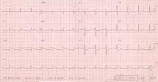
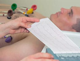
Also known as an ECG , the electrocardiogram is performed by placing electrodes on various parts of the patient's body. These electrodes, which are attached to the skin with clips or Velcro straps, are connected to the electrocardiograph through cables. In this way, the electrodes record the electrical signals of the heart and the electrocardiograph is responsible for measuring them, capturing them on a continuous roll of paper that constitutes the electrocardiogram.
There is a specific drawing that is considered a normal electrocardiogram: a P wave (corresponding to atrial depolarization), a QRS complex (ventricular depolarization) and a T wave (ventricular repolarization). This tracing reveals that there are no electrical alterations in the heartbeat. Other types of charts, however, can give a signal about a cardiac abnormality.
Through an electrocardiogram it is possible to detect an arterial blockage, an arrhythmia , ventricular hypertrophy or an electrolyte alteration, to name a few possibilities. That is why this exam is one of the most frequently used tools by cardiologists.
It is important to keep in mind, however, that not all heart diseases and disorders can be detected by an electrocardiogram, since it is limited to portraying electrical signals. There are pathologies that can only be discovered with a Doppler ultrasound ( Doppler ultrasound ) or other types of studies.
At a statistical level, it is known that many medical students are afraid of not being able to understand the basics of the electrocardiogram in order to read it correctly. This happens especially the first time they face one, and the fear is largely due to the pressure they know will exist the day they have to interpret it in front of a patient, without time to think or turn to a book.
 However, the problem does not lie in the complexity of the electrocardiogram but in the way in which university professors present it to their students: without explaining the origin of the waves that compose it, nor the possible anomalies in the case of disease. In other words, it is necessary to learn the theory but also have a context to properly understand a graph of this type, since it always comes from a real person, whose medical history we should also know to make an accurate judgment.
One of the tips to follow when faced with an electrocardiogram is to read it systematically, with the aim of finding one or more anomalies . This way of approaching interpretation is the same as in many other medical studies, since we should never quickly look for the points that catch our attention, but rather look at the entire document in order.
It is very important to know that the electrocardiogram looks different in each person, that is, both the image of normality and the opposite are particular to each person, and that is why they must be understood in context. To prepare a medical student adequately, it is necessary to teach him a large number of stereotypes , of possible normal patterns, so that he learns to dynamically adapt to the situation of each patient.
This brings us to the fundamental step before reading an electrocardiogram: knowing the patient. We must not forget that this record of his cardiac activity starts from him, and that is why it is necessary to know his state of health, his personality, his habits and his activities, which often alert us of a heart attack before the electrocardiogram.